breast


Expand All | Collapse All
Normal
- glands
- stroma
- intramammary lymph node
- signout
- Benign breast tissue
- No atypia, in situ or invasive carcinoma identified
Specimen types
- partial mastectomy = segmental mastectomy = lumpectomy = lump of breast fat
- total mastectomy = simple mastectomy = breast with skin and nipple
- skin sparing mastectomy: breast with nipple and areola, spares skin
- nipple sparing mastectomy = total skin sparing mastectomy: spares all of skin, nipple and areola
- radical mastectomy: includes axillary nodes and muscle
- modified radical mastectomy: includes fascia, +/- axillary node sampling
Clinical
- Early stage
- criteria: tumor <= 5 cm and <= 3 nodes
- stages: I, IIA, T2N1
- tx: surgery, sentinel lymph node
- caveats: dissection if 3+ positive SLNs or positive palpable node
- Locally advanced
- T3N0, stage IIIA-C = either tumor > 5 cm or > 3 node mets
- tx: neoadjuvant chemo, surgery, node dissection
- Metastatic = stage IV = contralateral node
- liver, pulmonary mets = even worse prognosis
- Hormones
- ER/PR status: dual positive > single positivity > all negative
- Her2 positive > negative due to Herceptin tx option
- Worse clinical factors: wt loss, poor performance status, LDH elevation, fast relapse
- Tumor burden markers: CA15-3, CA27.29, circulating tumor cells
Approach
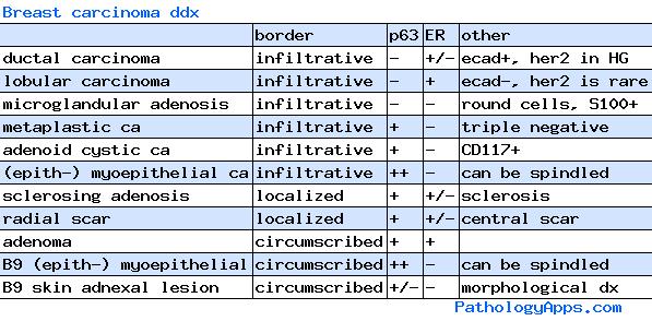
- epithelial lesions
- ductal
- lobular
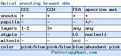
- other lesions: papillary, columnar cell, adenosis
- other carcinomas, metastasis
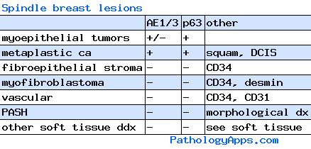
- stromal lesions
- infiltrate: heme, soft tissue
- fibroepithelial
- PASH
- clinical correlation
- calcifications
- does lesion explain mass
Ddx by categories
- intraductal lesions
- UDH
- ADH
- DCIS
- ductal carcinoma
- extension from lobular
- lobular lesions
- ALH
- LCIS
- lobular carcinoma
- extension from ductal
- papillary
- papilloma
- intraductal papilloma
- sclerosed papilloma
- papilloma with atypia
- papillary carcinoma
- intraductal papillary carcinoma
- encapsulated papillary carcinoma
- papilloma with DCIS
- papillary DCIS
- solid papillary carcinoma
- papilloma
- fibroepithelial lesions
- fibroadenomatoid change
- fibroadenoma
- hyalinized fibroadenoma
- phyllodes tumor
- juvenile fibroadenoma
- hamartoma
- columnar cell lesions
- benign epithelial proliferations
- sclerosing adenosis
- apocrine adenosis
- microglandular adenosis
- radial scar
- adenoma
- tubular adenoma
- lactating adenoma
- apocrine adenoma
- ductal adenoma
- sclerotic
- sclerosing adenosis
- radial scar
- scar
- fibrocystic change
- diabetic mastopathy
- sclerosed papilloma
- inflammatory
- mastitis
- diabetic mastopathy
- abscess
- fat necrosis
- biopsy site change
- granulomatous
- granulomatous mastitis
- biopsy site change
- sarcoidosis
- infectious
- reaction to implants
- cysts
- mucocele
- lactocele
- duct ectasia
- fibrocystic change
- apocrine cyst
- metaplasia
- mucin
- tubules
- other
Other neoplasms
- tubular carcinoma
- cribriform carcinoma
- medullary carcinoma
- atypical medullary carcinoma
- invasive carcinoma NST with medullary features
- apocrine carcinoma
- mucinous carcinoma
- signet ring carcinoma
- papillary carcinoma
- micropapillary carcinoma
- metaplastic carcinoma
- adenosquamous carcinoma
- squamous cell carcinoma
- spindle cell carcinoma
- myoepithelial carcinoma
- metaplastic carcinoma with mesenchymal differentiation
- mixed
- myoepithelial lesions
- myoepithelial hyperplasia
- collagenous spherulosis
- myoepithelial carcinoma
- epithelial myoepithelial lesions
- pleomorphic adenoma
- adenomyoepithelioma
- adenomyoepithelioma with carcinoma
- adenoid cystic carcinoma
- salivary gland tumor
- pleomorphic adenoma
- mucoepidermoid carcinoma
- adenoid cystic carcinoma
- acinic cell carcinoma
- skin adnexal tumor
- cylindroma
- clear cell hidradenoma
- sebaceous carcinoma
- other
- polymorphous carcinoma
- neuroendocrine (pure vs with neuroendocrine differentiation)
- secretory carcinoma
- oncocytic carcinoma
- lipid rich carcinoma
- glycogen rich clear cell carcinoma
- mesenchymal
- PASH
- granular cell tumor
- fibrous
- nodular fasciitis
- myofibroblastoma
- fibromatosis
- inflammatory myofibroblastic tumor
- solitary fibrous tumor
- vascular
- hemangioma
- angiomatosis
- atypical vascular lesions
- angiosarcoma
- peripheral nerve sheath
- neurofibroma
- schwannoma
- fat
- lipoma
- angiolipoma
- rhabdomyosarcoma
- osteosarcoma
- smooth muscle
- leiomyoma
- leiomyosarcoma
- hematolymphoid
- metastasis
- clinically diagnosed
- inflammatory carcinoma
- bilateral breast carcinoma
Grading (Nottingham score)
- glandular score
- wholistic, count only glands/cribriform with clear lumen surrounded by polarized cells
- 1: >75% glandular
- 2: 10-75% glandular
- 3: <10% glandular (lobular always get a 3)
- nuclear score
- worst area, compare to adjacent normal nuclei
- 1: normal-ish (< 1.5x size, minimal pleo, even chromatin, barely visible nucleoli)
- 2: cancer-ish (1.5-2x size, mild-mod pleo, visible but inconspicuous nucleoli)
- 3: ugly (> 2x, marked pleo, vesicular chromatin, prominent nucleoli)
- mitotic score
- worst area (usually leading edge), then choose 10 random fields within that area
- count only definite mits, ignore hyperchromasia/apotosis
- values below based on 10 40x fields with 0.55mm field diameter
- 1: <= 3
- 2: 4-7
- 3: >= 8
- use 40x
- overall grade
- 1: score 3-5
- 2: score 6-7
- 3: score 8-9
Hormone status
- ER and PR (nuclear stains, Allred system)
- intensity: 0-3
- 0: none
- 1: weak
- 2: moderatek
- 3: strong
- proportion: 0-5
- 0: none
- 1: <1%
- 2: 1-10%
- 3: 11-33%
- 4: 34-66%
- 5: >66%
- status: positive if total score 3+
- intensity: 0-3
- Her2 (membrane stain)
- negative (scores 0 and 1+): faint incomplete or < 10%
- 0: no staining or <= 10% faint incomplete staining
- 1+: > 10% faint incomplete staining
- equivocal (2+, send for FISH): weak-moderate complete staining > 10%, moderate-intense but incomplete, or <= 10% complete intense staining
- positive (3+): > 10% intense complete staining
- negative (scores 0 and 1+): faint incomplete or < 10%
- Her2 (FISH)
- negative: ratio < 1.8 or copy number < 4
- borderline (retest): ratio 1.8-2.2 or copy number 4-6
- borderline negative: ratio < 2
- borderline positive: ratio 2+
- positive: ratio > 2.2 or copy number > 6
TNM (useful for grossing)
- Tis: in situ
- T1: <= 20mm
- T1mi: <= 1mm (microinvasion)
- T1a: <= 5mm
- T1b: <= 10mm
- T1c: <= 20mm
- T2: <= 50mm
- T3: > 50mm
- T4: involvement of skin or chest wall
- T4a: chest wall (other than pectoralis muscle)
- T4b: skin ulceration, nodules, or edema (peau d'orange)
- T4c: both a and b
- T4d: inflammatory carcinoma
- N0: histologically negative, and...
- N0 (i-): IHC negative
- N0 (i+): IHC positive for isolated tumor clusters (<= 0.2mm and <= 200 cells)
- N0 (mol-): molecular negative
- N0 (mol+): molecular positive
- N1mi: micrometastases (tumor 0.2-2 mm or > 200 cells)
- N1a: macrometastases (tumor > 2mm)
- N2a: > 3 nodes, at least one macrometastasis
- N3a: >= 10 nodes, at least one macrometastasis
Staging
- 0: Tis
- IA: T1 or T1mi
- IB: T0-1, N1mi
- IIA
- T0-1, N1
- T2
- IIIA
- T0-2, N2
- T3, N1-2
- IIIB: T4, N0-2
- IIIC: N3
- IV: M1
CPT codes
- 88309: mastectomy with node dissection
- 88307: mastectomy, malignant lumpectomy, positive margin, sentinel lymph node (each), node dissection (lumped together)
- 88305: biopsy, benign lumpectomy, negative margin
- 88360: ER, PR, Her2
- 88342: stains
