liver
 Expand All
 | Collapse All
- segments
- zones
- periportal = zone 1 = around portal tract
- midlobular = zone 2
- centrilobular = zone 3 = around central vein
- portal tracts
- portal vein: ~5x size of artery or duct
- hepatic artery
- bile duct: always present. 10% of small tracts can lack it. Ductopenia if otherwise.
- parenchyma
- architecture: cords or plates of hepatocytes, 2-3 cells thick.
- hepatocytes
- sinusoids: lined by endothelial cells, Kupffer cells, stellate cells.
- bile canaliculi
- canals of Herring
- central vein
- aging changes: lipofuscin, atrophy
PatternsNon-neoplastic- inflammation
- metabolic
- biliary
- Explant
- cirrhosis with large regenerative nodules, ___ gram, clinical history of ___
- macrovesicular steatosis involving __ % of parenchyma
- other findings
- hilar margins appear patent
- negative for malignancy
- gallbladder findings
WHO neoplasms- Hepatocellular
- benign
- premalignant
- malignant
- Biliary
- benign
- premalignant
- malignant
- papillary neoplasm
- with intraepithelial neoplasia
- with invasive carcinoma
- mucinous cystic neoplasm
- with intraepithelial neoplasia
- with invasive carcinoma
- Uncertain origin
- calcifying nested epithelial stromal tumor
- carcinosarcoma
- combined hepatocellular-cholangiocarcinoma
- hepatoblastoma, mixed epithelial-mesenchymal
- malignant rhabdoid tumor
- Mesenchymal
- GCT
- neuroendocrine neoplasm
- lymphoma
- metastasis
Grading- HCC (descriptive)
- well differentiated: early lesions, usually < 2 cm
- moderately differentiated: 3+ cell thick trabeculae, looks similar to normal hepatocytes
- poorly differentiated: later lesions, pleomorphism
- undifferentiated: contains little cytoplasm, solid growth
- use highest grade found
- HCC (Edmonson and Steiner, not widely used)
- G1: rarely used
- G2: looks similar to normal hepatocytes
- G3: different from normal, but not overtly ugly
- G4: ugly
- use highest grade found
- biliary carcinoma
- G1: >95% glands
- G2: >=50% glands
- G3: >=5% glands
- G4: <5% glands
- Chronic hepatitis (Batts-Ludwig)
- 0: none
- 1: portal - patchy inflammation. Lobular - minimal necrosis.
- 2: portal - mild inflammation, in some or all tracts. Lobular - mild hepatocellular damage
- 3: portal - moderate inflammation in all portal tracts. Lobular - noticeable hepatocellular damage
- 4: portal - severe inflammation, bridging necrosis. Lobular - diffuse hepatocellular damage
- fibrosis/cirrhosis stage (Batts-Ludwig)
- 0: none
- 1: portal fibrosis
- 2: periportal fibrosis, rare portal-portal septa
- 3: septal fibrosis
- 4: cirrhosis (nodules)
- NAFLD activity score
- fat
- 0: minimal < 5%
- 1: mild <= 33%
- 2: moderate <= 66%
- 3: marked 67+
- balloon cells
- lobular inflammation
- 0: none
- 1: 1 foci per 20x field
- 2: 2-4 foci per 20x field
- 3: 5+ foci per 20x field
TNM (useful for grossing)- liver
- T1: solitary tumor
- T2: multiple tumors or vascular invasion
- T3a: multiple and >5cm
- T3b: portal/hepatic vein major branch involvement
- T4: adjacent organ (non-gallbladder) invasion or visceral peritoneum perforation
- biliary
- Tis: intraductal
- T1: solitary tumor
- T2a: vascular invasion
- T2b: multiple tumors
- T3: visceral peritoneum perforation or extrahepatic invasion
- T4: periductal invasion
Staging- liver
- I: T1
- II: T2
- IIIA: T3a
- IIIB: T3b
- IIIC: T4
- IVA: N1
- IVB: M1
- intrahepatic biliary
- I: T1
- II: T2
- III: T3
- IVA: T4 or N1
- IVB: M1
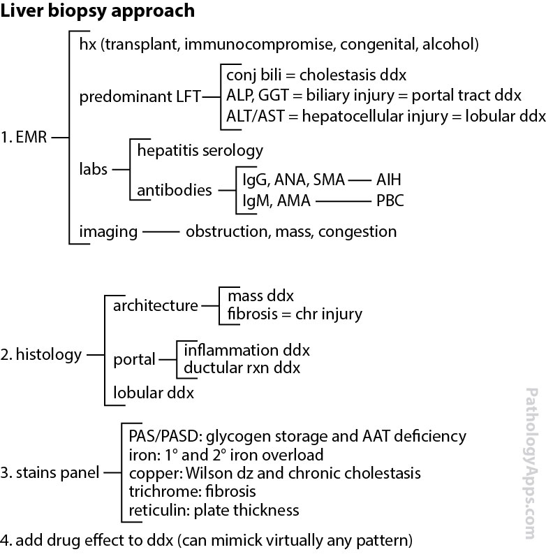
Stains- trichrome: fibrosis
- PASD: alpha1 antitrypsin deficiency globules
- iron: hemosiderosis
- copper: cholestasis, Wilson disease
- reticulin: plate thickness
|