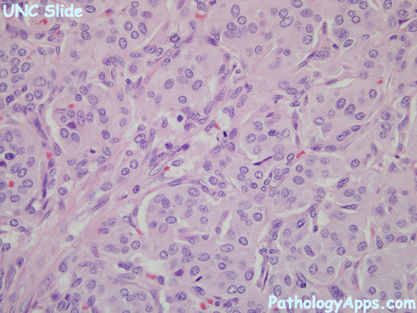
neuroendocrine neoplasm
 Expand All
 | Collapse All
WHO 2010 Grading and classification- neuroendocrine tumor G1
- mitosis < 2 per 10 hpf
- Ki67 <= 2%
- bland cytology, strong chromogranin and synaptophysin
- aka: well differentiated endocrine tumor, carcinoid tumor
- neuroendocrine tumor G2
- mitosis 2-20 per 10 hpf
- Ki67 2-20%
- bland cytology, strong chromogranin and synaptophysin
- aka: well differentiated endocrine carcinoma, atypical carcinoid tumor
- neuroendocrine carcinoma
- mitosis > 20 per 10 hpf
- Ki67 > 20%
- loss of chromogranin, retains synaptophysin
- atypia, necrosis
- types: small cell and large cell
- aka: NET G3, poorly differentiated NEC, small cell ca, large cell NEC
- mixed adenoneuroendocrine carcinoma
- both ductal adenoca and NEC, at least 30% of either
- squamous component is rare
- aka: mixed exocrine-endocrine carcinoma
- hyperplastic and preneoplastic lesions = tumor like lesions
- use higher grade re: mitosis vs Ki67
- requirements: 50 hpf for mitosis count, 500 cells for Ki67
- evidence support for stomach, duodenum and pancreas.
Stains- pos: synaptophysin, chromogranin
- rectal carcinoids: PAP+, PSA- (vs prostate PAP+, PSA+)
- lung (TTF1+, PAX8-) vs GI (TTF1-, PAX8+)
Hormones- nonfunctional
- serotonin
- gastrin
- glucagon
- insulin
- somatostatin
- VIPoma
Sign out- grade / classification
- mitosis count per 10 hpf
- Ki67
- margins
- TNM
|