meningioma
    Expand All
 | Collapse All
Clinical- most common intracranial tumor
- predominance in middle aged females
- associated with NF2 (22q)
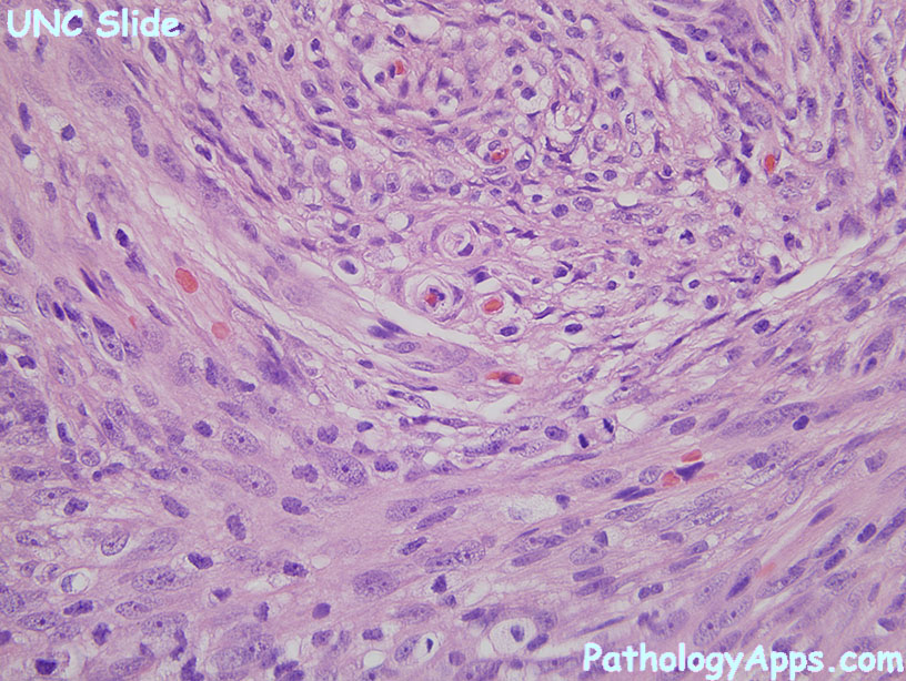
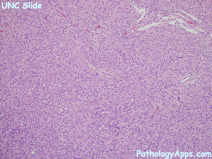
Grading, Subtypes- grade I, common
- meningothelial (oval cells, pseudonuclear inclusions)
- fibrous (collagen bundles)
- mixed (meningothelial + fibrous)
- grade I, others
- psammomatous (psammoma > tumor)
- angiomatous (vessels > tumor)
- microcystic (white spaces)
- secretory (PAS+ globules)
- lymphoplasmacyte-rich (inflammation > tumor)
- metaplastic (mesenchymal, myxoid, xanthomatous)
- grade II
- chordoid (pink cells in gray-blue mucinous matrix)
- clear cell (glycogen laden cells)
- atypical meningioma
- hypercellular with 5+ mitoses per 10 hpf
- alternatively, 3 of the following
- hypercellular
- sheet like
- necrosis
- 4 mitoses per 10 hpf
- small cells with high NC ratio
- prominent nucleoli
- grade III
- papillary (pseudopapillae, perivascular pseudorosettes)
- rhabdoid (eccentric nuclei, pink cytoplasm)
- anaplastic (20+ mitosis per 10 hpf, high grade cytology
Grading, other- brain invasion = grade II
Stains- positive: EMA (less so in atypical and anaplastic), vimentin, PR
- varies: S100
- secretory variant: CEA+
|