glomus tumor
     Expand All
 | Collapse All
Clinical- site: distal extremities, rarely in stomach
- size: < 1cm
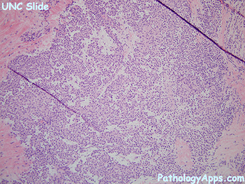
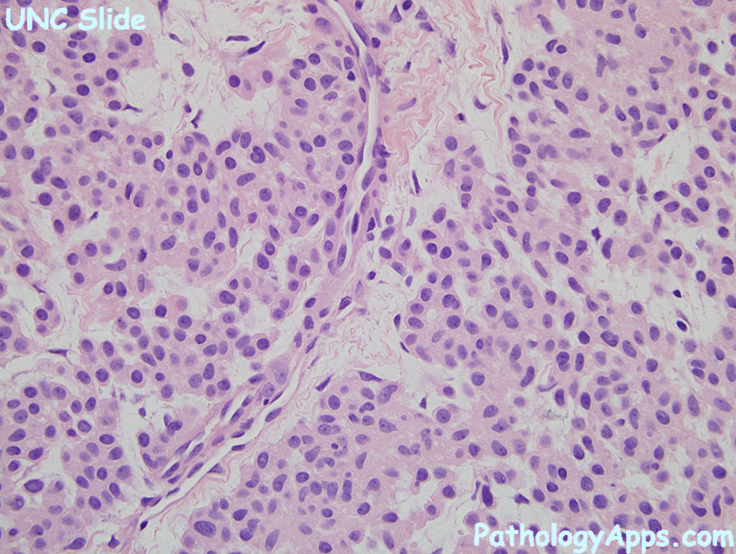
Histology- Components
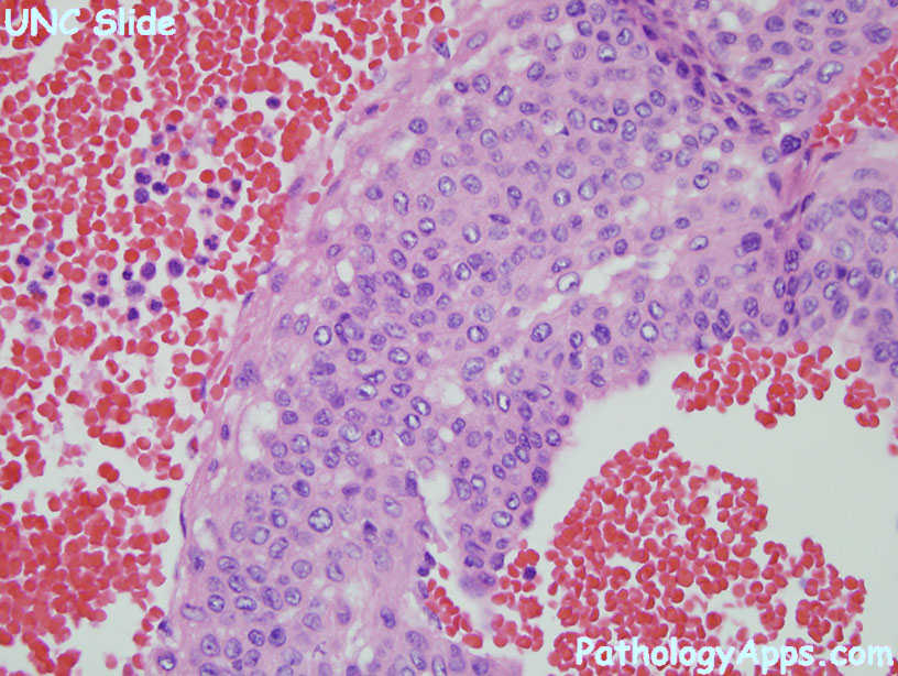
- glomus cells: small, round, central nuclei, surrounded by basal lamina
- vessels
- smooth muscle
- Subcategories
- solid glomus tumor
- 75%
- nests of glomus cells, capillaries
- glomangioma
- 20%
- small clusters of glomus cells, dilated veins
- glomangiomyoma
- nests of glomus cells and smooth muscle, staghorn clefts
Atypical variants- glomangiomatosis
- resembles diffuse angiomatosis
- but has glomus cell nodules in vascular walls
- symplastic glomus tumor
- degenerative atypia
- but no real atypia, necrosis or mitosis
- malignant glomus tumor
- size > 2cm and subfascial or visceral location
- atypical mitotic figures
- marked nuclear atypia
- glomus tumor of uncertain malignant potential
- has some but not all of the malignant features
Stains- positive: SMA, type IV collagen, h-caldesmon
- negative: desmin, CD34, CK, S100
|