acquired RCC


Expand All | Collapse All
Clinical
- ESRD on dialysis
Histology
- background ESRD: cysts, glomerulosclerosis, tubular atrophy, interstitial fibrosis
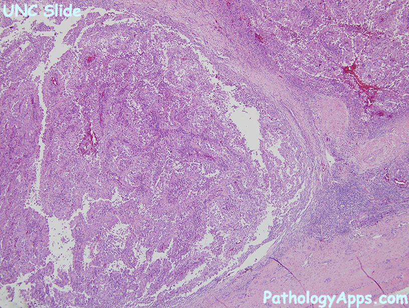
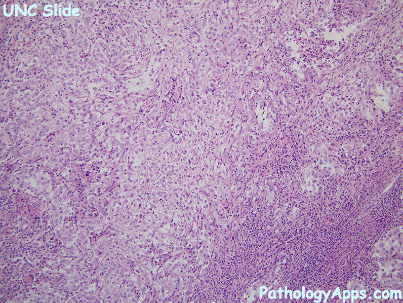
- architecture: solid, papillary, acinar, cribriform, tubulocystic
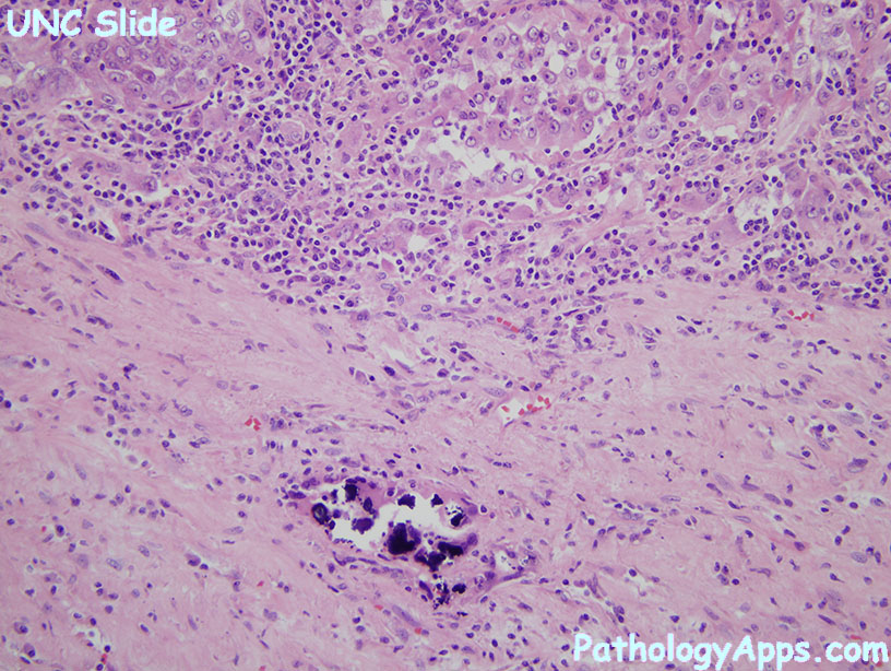
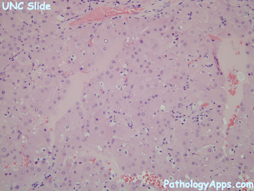
- cytology: pink vacuolated cytoplasm
- calcium oxalate crystals
Stains
- positive: CK, CD10, RCC, racemase
- negative: CK7 (at most focal)
