DFSP
       Expand All
 | Collapse All
Clinical- indurated plaques, nodules
- site: trunk, proximal extremities, head/neck
- superficial, low grade, locally aggressive (but rarely metastasize)
- high recurrence rate
- 10-15% progress to fibrosarcomatous DFSP
- tx: wide excision (preferably 2-3 cm margin), imatinib
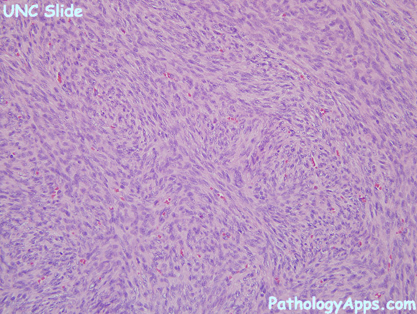
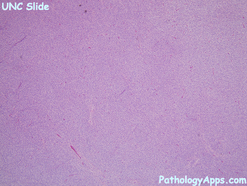
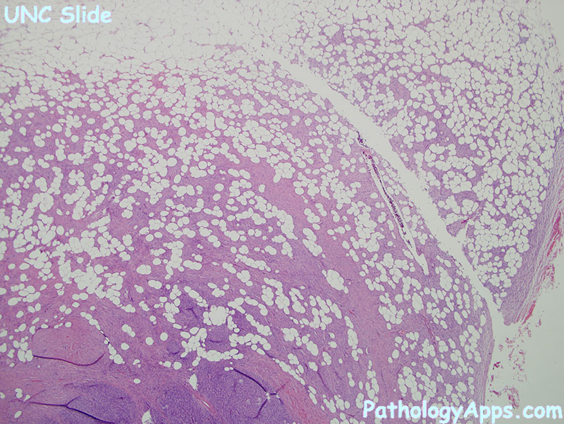
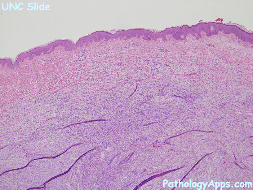
Histology- infiltrating dermis and subq, sparing epidermis
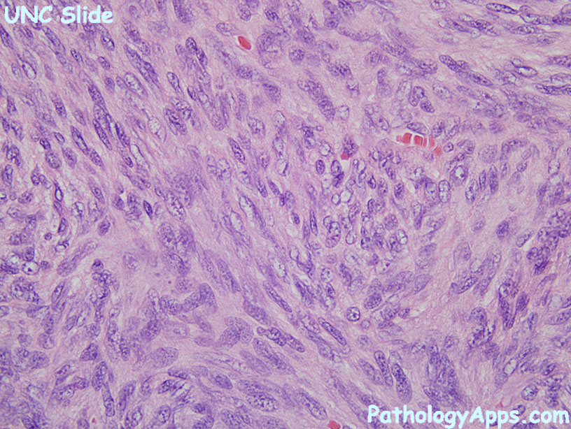
- densely hypercellular, spindle cells, storiform/whirling
- low grade cytology: minimal atypia or mitosis
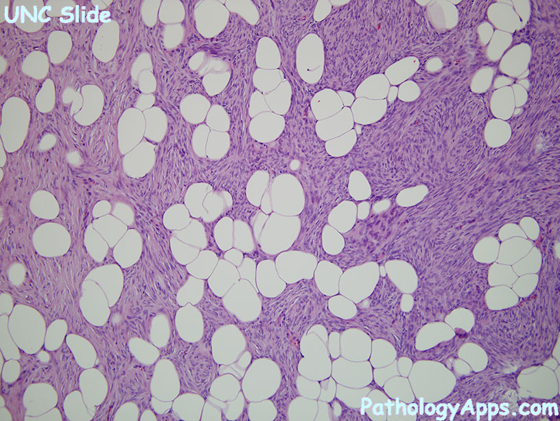
- honeycomb fat infiltration: tumor involves fibrous septa, interdigitating the fat cells
Variants- fibrosarcomatous DFSP: increased atypia and mitosis, reduced/loss of CD34, increased p53
- pigmented DFSP (Bednar tumor): has pigmented cells
- myxoid DFSP: myxoid, lots of vessels
- DFSP with myoid differentiation
- plaque like DFSP
- giant cell fibroblastoma = juvenile, multinucleated giant cells lining vasculature
Molecular- COL1A1-PDGFB fusion
- t(17;22)
Stains- positive: CD34
- negative: desmin, S100, CK
- can be weakly positive for EMA
- fibrosarcomatous DFSP: reduced/loss of CD34 in half, increased p53
- myoid differentiation stains SMA
|